While screen-film mammography has been proven to reduce the mortality of the disease, the screening method has its limitations. As Professor Keith Paulsen explains in this video published by the Thayer School of Engineering, doctors often find the images created by mammography difficult to read due to “tissue overlap”—a type of distortion that occurs when a complex, 3D object is rendered as a 2D image:
Approved for use by the FDA in 2011, tomosythesis collects imaging data much like an MRI scan, except that the X-ray generator rotates around the patient and then mathematically reconstructs this data into a 3D model. Currently, MD/PhD student Kelly Michaelsen and Professor Venkataramanan Krishnaswamy are developing a screening system that combines near infrared spectral tomography (NIRST) with the high-resolution 3D structural information of breast tomosythesis (BTS) into a singular breast-cancer detection method. This research is being conducted with both Hologic, Inc—the commercial leader in the development of breast tomosythesis (BTS) technology—and the University of Massachusetts Medical School.

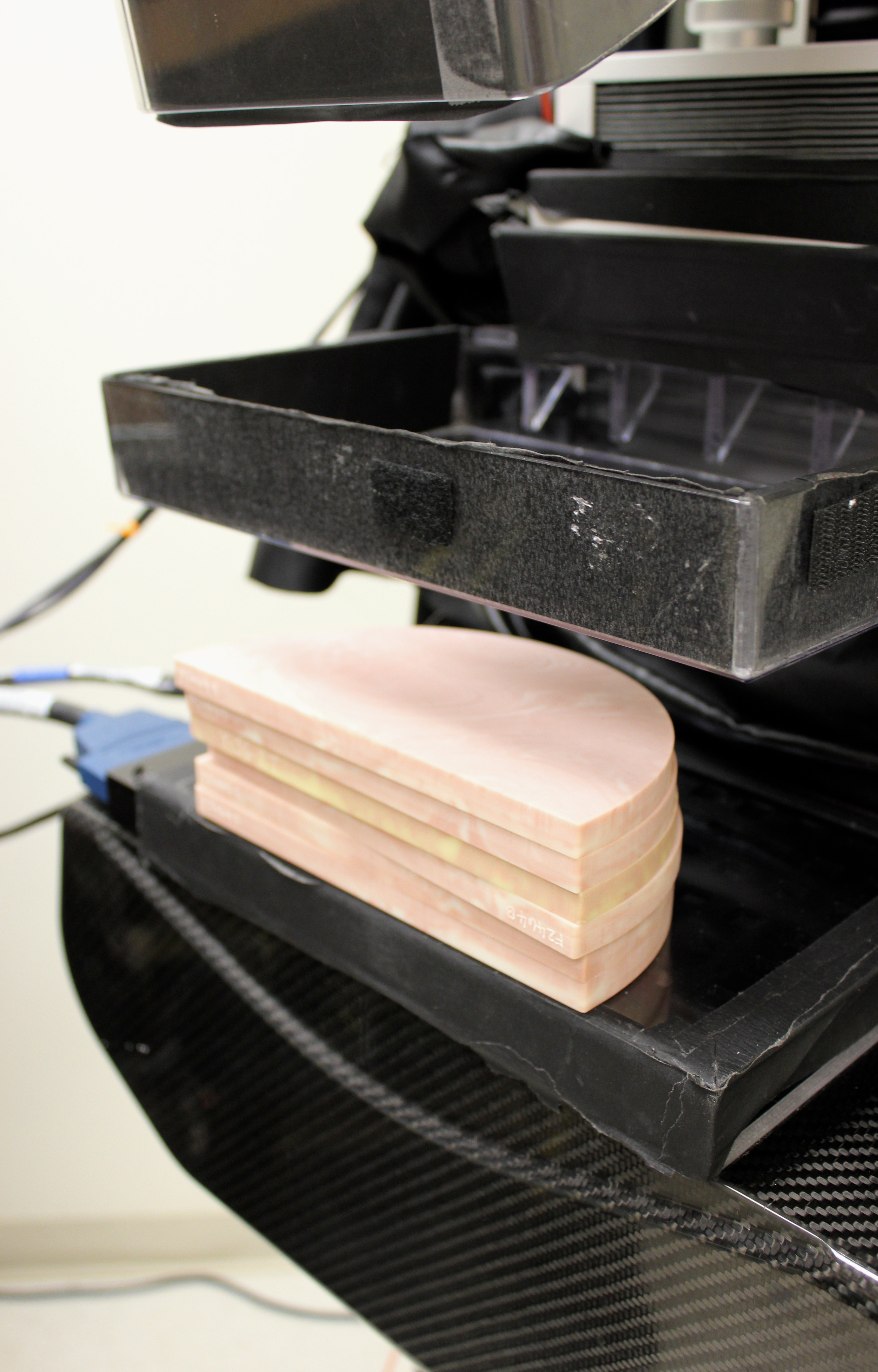
The first prototype of this imaging system is currently being tested at the Dartmouth Hitchcock Medical Center (DHMC). After calibrating its components on a series of tissue phantoms, Michaelsen and her advisor started conducting pre-clinical imaging trials on patients at DHMC. With the data collected from these trials, the research group identified aspects of the system that could be improved, and began constructing a second-generation prototype of the imaging machine. Once this second-generation prototype is completed, the original machine will be moved to the University of Massachusetts Medical School where researchers will collect clinical data in another series of pre-clinical trials.
“In 2010, the fatality rate for females diagnosed with breast cancer in the US was just over 19 percent,” says Michaelsen. “The combined imaging system that our lab is developing is aimed at decreasing the number of women who go through invasive biopsy procedures. Through improving the early stages of breast cancer detection, we hope to decrease the fatality rate of this disease.”

The research being conducted at all of the institutions involved in this project has been made possible by a funding opportunity provided by the National Institute of Health (NIH) and its National Cancer Institute (NCI) that seeks to develop new in vivo imaging systems. The partnership between Dartmouth, the University of Massachusetts, and Hologic, Inc aims to integrate the two systems into a singular detection method, and to establish the clinical potential of this combined imaging approach.

