Located on the second floor of the Dartmouth Hitchcock Medical Center (DHMC), the Advanced Imaging Center houses several imaging devices used by the Optics in Medicine Laboratory. Currently, graduate student Michael Mastanduno is testing a prototype that combines Magnetic Resonance Imaging (MRI) with Optical Tomography into a singular imaging device. This has the potential to significantly improve the accuracy of non-invasive breast-cancer imaging, leading to better patient care.

This summer, graduate student Fadi El-Ghussein started working on and upgrade to this system’s instrumentation. El-Ghussein’s input enabled the research team to finalize a prototype of this non-invasive imaging machine and to begin testing it on humans in a clinical setting. To expedite the testing process, the research team is moving the imaging system to Xijing Military Hospital in Xi’an, China for two months with Associate Professor Shudong Jiang.
“Due to our remote location in the Upper Valley, we’re only able to image a small number of patients each year,” explains Mastanduno. “However, in Xi’an, our team will be able to test the imaging machine on two patients a day. Hopefully, the images gathered by our team in the Xi’an clinical tests will provide the data necessary to have the efficacy of the technology fully evaluated by 2012.

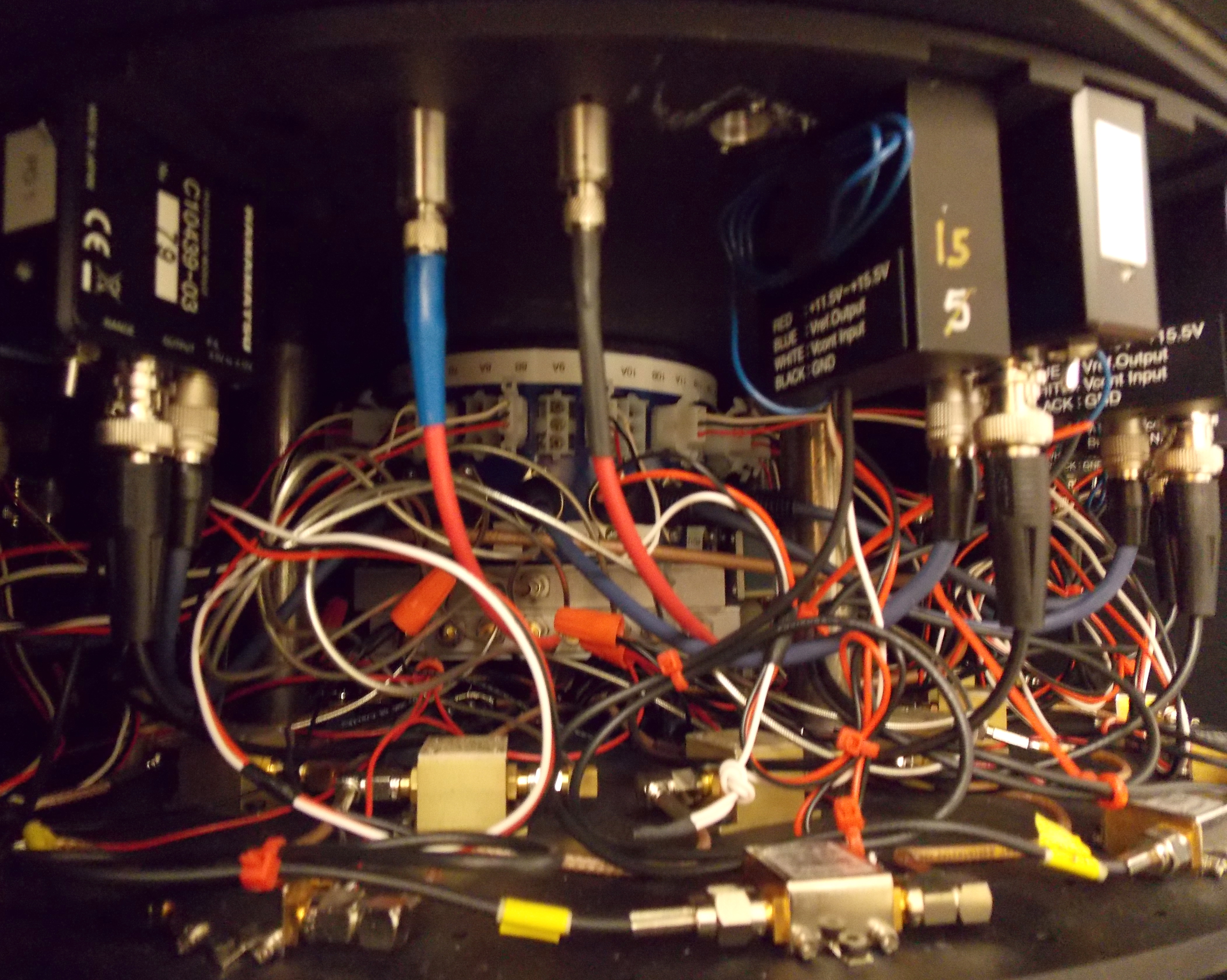
The instrumentation built by El-Ghussein enables the imaging machine to deliver optical light to the MRI scanner, and then to collect this data from either a tissue phantom, or a patient. While the components used by El-Ghussein to build this device are commonplace in clinical imaging systems, this machine is unique in that it allows both the MRI scanner and the machine’s optical components to independently collect data, and then feeds this information into a computer where it is mathematically reconstructed into a detailed 3D model.
“The instruments that I’ve developed over the last few months use both Photo Multiplier Tubes (PMT) and Photo Diodes (PD) to collect the data from the machine’s fiber-optic components. This data is then mathematically reconstructed into a tissue model, and is then combined with the magnetic data gathered through the MRI scan,” explains El-Ghussein. “I’m very happy with the data that our lab has collected so far in Upper Valley, and both Mike and I look forward to traveling to China to conduct clinical trials at the Xijing Military Hospital.”

To enable the imaging system to simultaneously collect MRI and tomographic data, Mastanduno engineered the machine’s cradle so that its optical fibers connect directly with the patient’s skin. Housed beneath the MRI’s breast coil, this cradle guides the fiber optic cables to the patient’s breast, and once each cable is firmly connected with the organ’s surface, the machine starts to collect imaging data.
“The great thing about this machine is that that it’s entirely non-invasive,” says Mastanduno. “While the patient might feel a slight compression as the optical cables connect with her breast or a warming sensation as the MRI machine gathers magnetic data, our lab’s imaging systems do not rely upon surgical procedures to collect data.”