Co-directors: Bruce Stanton, PhD, Professor of Microbiology and Immunology; George O’Toole, PhD, Professor of Microbiology and Immunology
Location: The ACBC is located in the Remsen Building (rooms 504 and 519) on the Hanover campus of the Geisel School of Medicine.
Staff: Roxanna Barnaby, Amanda Nymon
Contact: Bruce Stanton (bas@dartmouth.edu)
Description: The mission of the Airway Cell Biology Core (ACBC) is to advance basic science, as well as translational and discovery studies relevant to interactions of microbes with host epithelial cells from the airway, provide expertise using a co-culture platform of bacterial biofilms on human epithelial cells (including the Dartmouth Co-Culture Assay), and provide a range of airway cell lines, including primary human airway epithelial cells, 3D organoids and 2D organoid cultures from cystic fibrosis (CF) and non-CF donors and mice, and to support studies assessing host-microbe interactions relevant to the chronic infections associated with the CF airway. The ACBC also assists in the development of new cell lines and other methodologies to advance the science of the RDP members and Pilot Project awardees.
ACBC services include support for the following:
- Airway host-microbe co-culture studies. The ACBC has implemented 3D organoids and 2D organoid-derived monolayers from mice and humans, leveraged its expertise in co-culture models to study interactions between these models and lung bacteria, initiated the Lung-On-A-Chip model, and characterized bacterial isolates obtained from the lungs of CF and non-CF children and from endoscopic aspirates (CTRC).
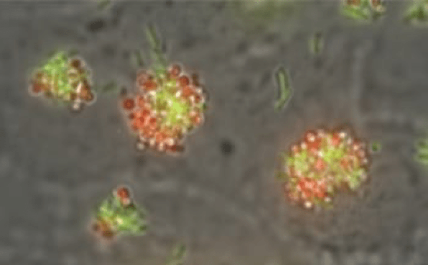
- Imaging studies of airway epithelial cells and bacterial biofilms. The core has a Keyence BZ800 microscope and access to several confocal microscopes at Dartmouth.
- Expertise in single cell-RNA-sequencing, other molecular techniques, and characterization of airway organoids.
- Functional studies including forskolin-induced swelling assays (FIS) of 3D GI organoids, and electrophysiological studies of 2D organoid monolayers.
- Studies on mouse models to study GI disease in CF.
- Access to lung bacterial and fungal strains.
- Coordination with industry to develop new multi-organ therapeutics relevant to CF.
- Training in the use of lung microbe-host co-culture organoid models and mouse models.
Major Equipment in the Airway Cell Biology Core:
| Equipment | Quantity |
|---|---|
| Easy Mount Diffusion Chamber and Clamp for Ussing studies | 6 |
| Beckman Coulter Z1 Particle Counter | 1 |
| BioRad TC10-automatic cell counter | 1 |
| Nanodrop ND-1000 Spectrophotometer | 1 |
| Agilent 2100 Bioanalyzer | 1 |
| NanoSight NS300 | 1 |
| A hood for virus and mouse work | 1 |
| Emulate Zoe and the Orb (Lung on a Chip) | 1 |
| Keyence BZ-X800 fluorescence microscope for biofilm studies | 1 |
Keyence BZ-X800 epi fluorescence microscope (filter cube sets):
| DAPI, GFP, TRITC and Texas Red |
| DAPI: Excite 360/40, emission: 460/50 |
| DAPI-V for sectioning: ex: 395/25 em:460/50 |
| GFP: ex 470/40, em 525/50 |
| TRITC: ex 545/25, em 605/70 |
| Texas red: ex 560/40, em 630/75 |