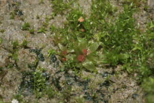
Physcomitrella patens is a moss that is used as the model organism for this experiment. It’s high plasticity and known genome make it perfect for Magdalena Benzanilla’s work (Source: Wikimedia Commons).
Recent research using moss may give more insight into how the components of the cytoskeleton communicate with each other. The cytoskeleton, a supportive, intracellular structure, is integral to cellular function. Composed of microtubules, actin filaments, and intermediate filaments, the cytoskeleton transports proteins and maintains cellular integrity. However, the mechanisms it uses to carry out these functions are not clear. At a special Dartmouth biology seminar this past week, Magdalena Benzanilla, a biology professor at the University of Massachusetts Amherst, presented research that clears up interactions between actin filaments and microtubules in Physcomitrella patens, a moss.
According to Benzanilla, Physcomitrella is advantageous for research because its entire genome has been sequenced. Its cells can be lysed – emptied of their contents – and developed from the spore phase. In the spore phase, Physcomitrella has high plasticity, meaning that it is resilient and flexible, and creates a filamentous network through mitosis, or cellular replication. The growth can occur symmetrically or asymmetrically, and it was found that microtubules and actin filaments play a crucial role in the direction of growth. Benzanilla found that they interact with the help of myosin VIII, a protein that aids actin filament motility, which gathers at the side of the cell where growth takes place and where the cell plate is formed. In order to fully divide the cell must create a cell plate. After anaphase, where the two sets of chromosomes move apart in the nucleus, the microtubules use myosin VIII to guide them in creating the cell plate.
Another protein observed was formin, which is used in degradation and regeneration of microtubules. In order to more fully characterize the importance of the proteins, Benzanilla performed experiments with cells that didn’t have the formin or myosin VIII genes. The resulting data further emphasizes the importance of both these genes within the cell.
Benzanilla’s current hypothesis is that formin plays a role in guiding the components of the cytoskeleton for cell division. When the formin gene is knocked out, or removed from the genome, the microtubules and actin filaments lose specific direction but generally still grow in the appropriate direction, albeit at a slower less concentrated rate. Less area is covered when myosin VIII is knocked out, but no other notable effects were observed.
More research is needed to confirm hypotheses, but so far Magdalena Benzanilla’s work confirms that formin and myosin VIII are both crucial in cell growth. Myosin VIII completes mitosis by designating where the cell plate is to form and guides cell growth directionality. Formin acts as a connecting factor between microtubules and actin filaments when cell growth occurs. Future research is needed to further shed light on the mechanisms of the cytoskeleton.