Dev Kapadia 23′
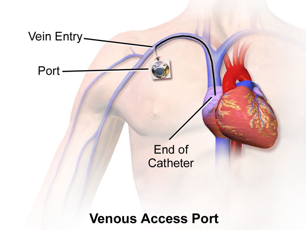
Figure 1:
This image depicts the location of a central venous catheter. Because of its more central location in the vascular system, it is far more invasive than needles administered at peripheral sites in the arm.
Source: Wikimedia
A critical part of medical procedures is gaining vascular access – opening up a patient’s blood vessels to either draw blood or administer medication. Such operations are extremely common as medical professionals in the United States perform over one billion vascular access procedures every year.1 Peripheral vessels, veins and arteries not located in the chest or abdomen, are of particular interest as they can be accessed far less invasively than those of the central vascular system. For this reason, over 90% of hospitalized patients receive a peripheral vascular access procedure at one point during their visit.1
Although these procedures are vital, the two choice methods of access, needles and catheters, each have major drawbacks. While needles are designed to be less invasive and would be safer option if performed perfectly, many medical professionals require several attempts to find the target vessels. As a result, sticking the patient multiple times can become increasingly unsafe as treatment is delayed, bleeding can become worse, risk of infection can increase, and vessels and surrounding tissues are more damaged as a result from several punctures.2 In treating pediatric, elderly, chronically ill, or trauma patients with small, twisted, or collapsed blood vessels that are even harder to access than those of healthy patients, medical professionals often need at least five attempts until a successful stick is administered, resulting in a first-stick accuracy rate of under 50 percent.3 Central venous catheters may be used instead to attempt to protect patients from risks associated with multiple needle sticks. However, these catheters are still far more invasive, more labor-intensive, and more likely to cause blood clots in addition to other blood-related issues compared with one to two needle sticks.2
To increase needle stick accuracy, engineers at Rutgers University have developed an image-guided robotic device to navigate needles to peripheral blood vessels. The device uses machine learning with near-infrared and ultrasound imaging to build a 3D image of the arm. This allows the device to distinguish target blood vessels from the surrounding environment, estimate the depth, and the follow the target vessels using motion tracking. The device can be used to insert both needles and catheters, draw blood, or deliver fluids and drugs to the bloodstream.3
Not only can the device successfully direct needles and catheters to the vessels, it has performed better than medical professionals during test runs. In the studies conducted by the Rutgers engineers, the device had an average of 0.3 failed access attempts per patient compared with 1.8 failed access attempts per patient for the human control group. Furthermore, the device had an 88% first-stick success rate by compared with a 53% first-stick success rate for the human control group. The Rutgers team found that the improvement was even more pronounced when the target vessel was small, twisted, or collapsed.4
While the device is currently designed for human procedures, the team claims that the device can be adjusted for peripheral vascular access in rats. This will make conducting drug testing clinical trials for pharmaceutical and biotech companies significantly more efficient. Along these same lines, the immediate next steps for the team will be to conduct further tests of the device to more subjects with blood vessels that are normal and difficult to access.3
References:
[1] Lamperti, M., & Pittiruti, M. (2013). Difficult peripheral veins: turn on the lights. British Journal of Anesthesia , 110(6), 888–891. doi: https://doi.org/10.1093/bja/aet078
[2] Radiological Society of North America. (2019, March 5). Vascular Access Procedures. Retrieved March 25, 2020, from https://www.radiologyinfo.org/en/info.cfm?pg=vasc_access
[3] Rutgers University. (2020, March 4). Robot uses artificial intelligence and imaging to draw blood: Engineers create device that can also insert catheters. ScienceDaily. Retrieved March 25, 2020 from www.sciencedaily.com/releases/2020/03/200304141540.htm
[4] Chen, A.I., Balter, M.L., Maguire, T.J. et al. (2020). Deep learning robotic guidance for autonomous vascular access. Nat Mach Intell 2, 104–115. https://doi.org/10.1038/s42256-020-0148-7