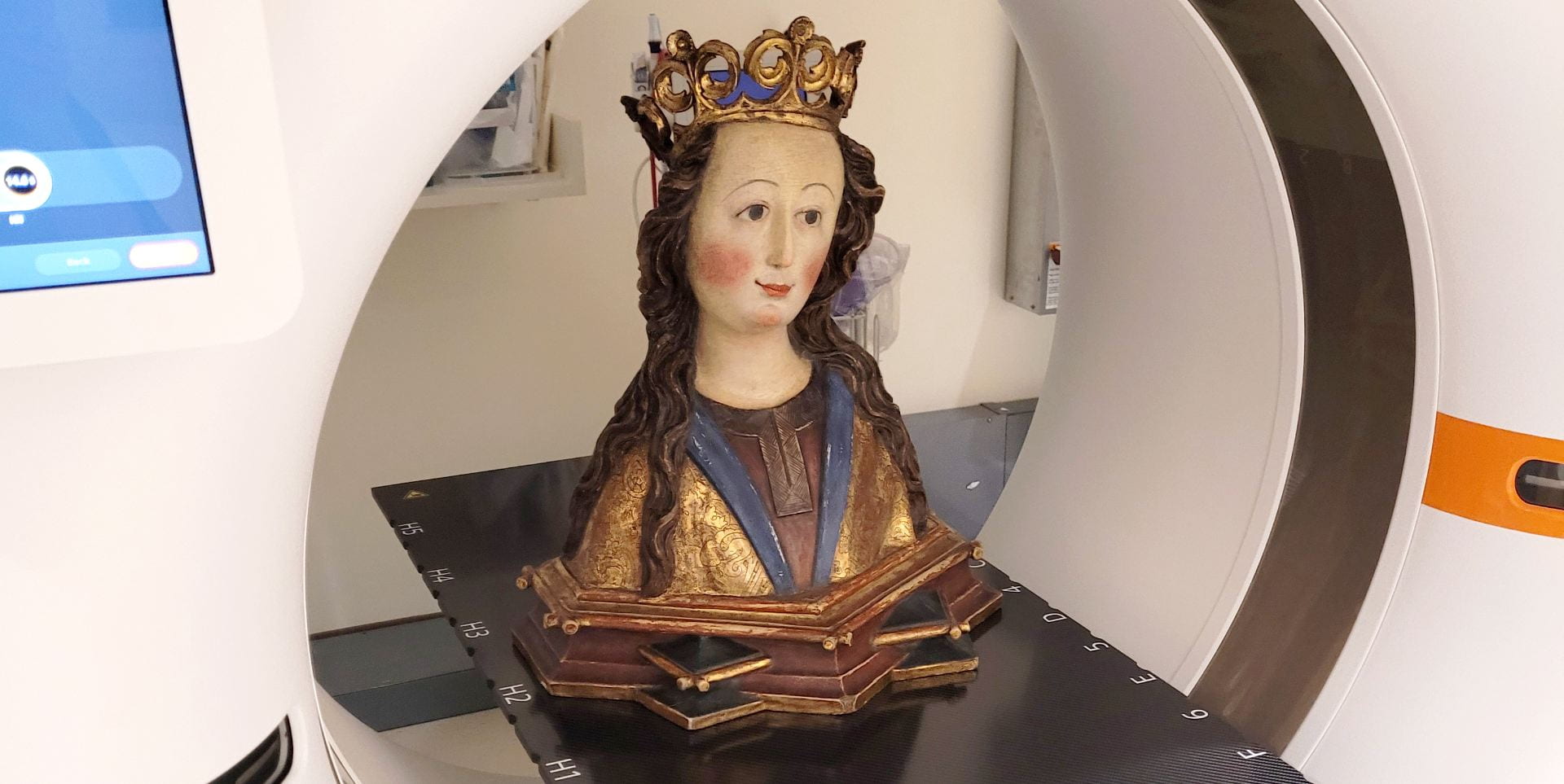
In the winter of 2022, the Hood acquired its first medieval reliquary. Purchased with the support of a grant from the Henry Moore Foundation, this sculpture filled an important gap in the Hood’s collection of medieval and Renaissance sculpture. Popular among Christians throughout the Middle Ages, reliquaries were made as containers for the bones of saints. These sculptures honored the holy person, created a focus for prayer, and served as a conduit between Heaven and Earth. Relics often motivated pilgrimages, which might draw worshippers from a great distance to pay their respects to a saint. Reliquaries, which could be made of wood, as in this case, or precious metal, offered proper, honorific housing for these important remains of a saint. Prior to this purchase, the museum did not have a reliquary in its collection, despite the significance of this sculpture type in the Middle Ages. While reliquaries could take many different shapes, from simple boxes to detailed body parts, the Hood’s new reliquary is in the form of a woman’s upper body.

Carved around 1500 in the region of Swabia—what is today southern Germany—the sculpture is made of wood and retains much of its original paint. The depicted woman wears a crown and has long, gently curling hair. Smiling slightly, the woman stares straight ahead, with her head tilted at an angle. She wears a robe with incised patterning in gold, blue, and red. Carefully painted, much of the original detail remains intact, including her individual eyelashes, distinguished by single brushstrokes. The base of the reliquary features offset and interlocking rectangles, an architectural motif common in Gothic decoration in southern Germany and northern Austria at the turn of the sixteenth century.
This reliquary would likely have once stood on an altar, the central focus of Christian rituals in a church. There, the sculpture would have enabled medieval audiences to reflect on the saint and her exemplary life. Although some saints are relatively simple for modern audiences to distinguish, others are more difficult to identify. Often a saint can be named by their association with an attribute, a distinguishing object or accompanying animal, such as Saint Jerome and the lion. Beyond her crown, however, this saint lacks a distinctive attribute. While her specific identity remains unknown, popularly worshipped women saints during the Middle Ages included Catherine, Barbara, and Margaret.
When the reliquary arrived in the museum, little was known of its history. Although this object’s provenance can be traced back to the beginning of the twentieth century, its makers and original placement remained unknown. Additionally, it was not certain whether the sculpture still contained its relic. Conservators often carry out scientific analysis, called “technical examination,” of materials and manufacture to study an object and its origins., The Hood, however, does not have conservators on staff. As a result, the quest to find out more about this reliquary led us to look for campus partners who had expertise in non-invasive examination techniques, which would not require taking samples from the object. One traditional method for studying the structure of wooden sculpture is X-ray imaging. This method allows researchers to see a cross-section of a work of art, much as it enables doctors to examine a body part. Increasingly, however, conservators and researchers are turning toward newer technology such as Computed Tomography (CT scans) to study works of art. CT scans bring together X-rays with digital imaging to create more detailed internal views of the body. In this case, CT scanning promised a better understanding of the structure of our reliquary bust.

Luckily, Dartmouth is home to both world class arts institutions and medical facilities. Staff at Dartmouth-Hitchcock Medical Center and faculty at Geisel School of Medicine were enthusiastic about applying their research methodologies to our medieval reliquary. Dr. P. Jack Hoopes, Professor of Surgery and Radiation Oncology and Dr. David Gladstone, Professor of Medicine at Geisel School of Medicine booked an appointment for our “patient” at the hospital. Together with Ashley Offill, Associate Curator of Collections, and Lauren Silverson, Registrar at the Hood, I brought the reliquary to Dartmouth-Hitchcock just a short drive down the road. In Radiation Oncology, Dr. Hoopes and Dr. Gladstone led the scanning of the reliquary, much as they would of any patient. While she was a good deal older and much quieter than the typical visitor to the hospital, our reliquary bust underwent several typical scans at varying centimeter slice thicknesses. In the five hundred years since she was made, this reliquary was now the beneficiary of the latest medical technology.
Her insides had a great deal of information to offer. First, the scan showed that there as a cavity in the top of the head of the reliquary, drilled straight down in the center of her cranium. While now empty, this space was possibly the site where the relics were originally housed. This placement in the head is in keeping with the location of relics in many bust-shaped reliquaries from this period. Associated with the body’s power, the head suggested the saint’s authority and gave the represented figure a physical presence. With individualized facial expressions and details, the head reliquary was life-like, a sensibility that would be augmented by the placement of the holy relic. Additionally, the CT scanner identified the remains of five nails in the back of the base, suggesting the sculpture may have been secured to a possible backing. These nails, an unusual discovery, may have been used to attach the reliquary to a backing or to keep it in place on an altar, potentially attaching it to a larger altarpiece ensemble that would have been a focus for Christian worship. It is also possible that the nails were added at a later point, after the reliquary was removed from its original location and installed elsewhere.

Additionally, imaging revealed that the reliquary was made of different blocks of wood put together and then covered in paint to disguise the joins. The shoulders and base are made from separate blocks held by wooden pegs and metal nails. A large knot in the wood, running through the reliquary’s chest was immediately visible on the scans, though hidden inside the sculpture. Finished with delicate painting, the reliquary would have served to honor a saint and offer a focus for its religious community.
Although very finely painted and impressive on the exterior, the interior composition suggests that this sculpture was part of a workshop’s serial production. This compositive structure of imperfect wood would have been relatively cheap and easy to produce. A kind of “off the rack” carving, the reliquary could then be personalized by a church with the insertion of the local relic. With no specific attributes, the reliquary could become any number of women saints, depending on which relic the church owned, whether a widely venerated one like Saint Catherine or a more localized figure such as Saint Hedwig. Sculptors in southern Germany were not alone in mass-producing reliquaries; carvers near the city of Cologne in western Germany had created reliquaries of St. Ursula in large numbers in the early fourteenth century.

The CT-scanner was further able to detect the densities of the materials used to make the sculpture. Two different woods registered on the scanner: the chest of the bust is slightly lower in density than the rest of the reliquary, which is all carved from the same wood. One future avenue for research could be to gather sufficient data to determine wood type based on density readings. Nails used to secure the blocks of wood on the shoulders and base are made of a copper alloy, consistent with the type of metal nails commonly used in this period. Underneath the paint, we also discovered a chalk layer ground, which is consistent with painting techniques in Germany at the turn of the sixteenth century.
After her brief trip to the hospital, the reliquary has returned to the museum. She plays an active part in teaching at the museum and will be featured in an upcoming exhibition of medieval and Renaissance sculpture. We are excited by the results of the CT scans, our first step in looking at this work from the inside and out. Without the assistance of staff and doctors at Dartmouth-Hitchcock and Geisel School of Medicine, none of this research would be possible, and we look forward to future opportunities to bring together the arts and sciences at Dartmouth!
This post was authored by: Elizabeth Rice Mattison, Andrew W. Mellon Associate Curator of Academic Programming

ABOUT THE AUTHOR
Elizabeth (Beth) Rice Mattison joined the staff in July 2021 as the Andrew W. Mellon Assistant Curator of Academic Programming. She serves as the liaison to Dartmouth faculty and facilitates the integration of the museum’s collection across the College’s curriculum. A specialist in medieval and early modern art, especially in northern Europe, she publishes widely in museum publications and scholarly journals including Gesta, Metropolitan Museum Journal, and Burlington Magazine. At Dartmouth, she’s committed to engaging diverse audiences with objects to elicit critical thinking and foster transformative encounters with art. Beth earned a PhD in art history from the University of Toronto and an MA and BA in the history of art from Yale University. She has held positions at several institutions, including the Centre for Renaissance and Reformation Studies at the University of Toronto; Musée du Louvre; John and Mable Ringling Museum of Art; and Yale University Art Gallery.

Comments are closed.