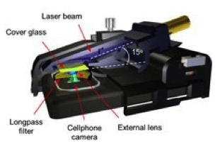
On March 5, Qingshan Wei, Postdoctoral Scholar in Electrical and Bio-engineering at UCLA, gave a seminar on his research into developing mobile phone-based systems for imaging, sensing, and diagnostics. Although the first compound microscope was invented in 1595, the past three decades have seen rapid advances in imaging technology. However, while specificity, contrast, and resolution have improved significantly, factors such as portability, cost-effectiveness, and simplicity have not improved recently.

The mobile phone-based microscope that Wei and colleagues in the Ozcan Lab at UCLA developed in 2010 was portable, cost-effective, easy to use, had high sample throughput, and could be mass-produced.
Courtesy of the Ozcan Research Group of UCLA Engineering
In addition, three billion people around the world are currently medically underserved, despite these technological advances. Wei focuses his research on field detection and point-of-care applications of his mobile phone-based systems in order to eventually bridge the gap between the laboratory bench and applications for large populations.
The mobile phone-based microscope that Wei and colleagues in the Ozcan Lab at UCLA developed in 2010 was portable, cost-effective, easy to use, had high sample throughput, and could be mass-produced. In addition, the phone-based system combined functionalities such as image acquisition, storage, analysis, and sharing into a single platform. Furthermore, the computational power of mobile phones has recently been catching up to PC’s. Although reductions in pixel size and expansion of pixel number have increased resolution, the smallest detection size of the mobile phone-based microscope is 1 micron, or 10-6meters.
Seeking to push the limits of phone-based imaging to nanoscale sensitivity, Wei and colleagues used a very oblique (75°) angle of illumination from a laser diode to minimize background noise in images, allowing their phone-based system to detect distances down to 100 nm (0.1 micron) as of 2013. With this level of sensitivity, Wei was able to detect individual virus particles using fluorescent antibody tags.
This has potential applications in diagnosis of severe viral infections such as HIV. One of the major current methods of HIV diagnosis is indirectly measuring immune function, but the actual number of virus particles provide a better picture regarding the progress of the infection. Thus, this mobile phone-based nanoimaging could potentially help detect HIV earlier when there is a smaller viral load.
This new phone-based system was found to have comparable sensitivity to a 100x confocal microscope, despite its much smaller size. Wei’s system was also able to detect individual DNA molecules to within hundreds of base pairs (bp) for DNA molecules 10,000 bp and longer. Measuring the length of DNA molecules could be used to analyze duplications or deletions of genes, which would alter DNA length. Such mutations have been associated with numerous genetic disorders, suggesting important applications for this DNA length measurement system.
Researchers in the Ozcan lab also developed cell phone-based biomarker and toxin detection systems, which could detect mercury levels down to 3.5 parts per billion (ppb). For context, the WHO recommends no more than 6 ppb of mercury and the EPA recommends no more than 2 ppb.
Moving away from the mobile phone platform, the third major project Wei presented was lens-free on-chip imaging. This type of imaging extends recent “lab-on-a-chip designs” to include imaging as well, further increasing cost-effectiveness and portability compared to the mobile-phone based systems. Using interference patterns formed by light moving through an aperture, this system could digitally reconstruct the actual image, nearing a 10X microscope in sensitivity. Via machine learning algorithms, the on-chip imaging system was able to distinguish between two types of medically important immune cells with 95% accuracy after silver and gold nanoparticle labeling.
Wei hopes to pursue further research into portable imaging technology, on-chip imaging, microfluidics, and detection of biologically important molecules and particles. His research will continue to find direct application in personalized healthcare and point-of-care diagnostics.

Leave a Reply