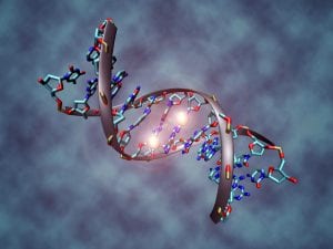
An artist’s rendition of a DNA segment showing two methyl groups, represented as glowing orbs attached to specific base pairs. DNA methylation plays an essential role in gene regulation during development or in the presence of environmental stimuli. (Source: Wikimedia Commons, Christoph Bock)
The ongoing debate between nature and nurture clashes in the field of epigenetics, the study of how the environment changes gene expression. DNA methylation, a primary mechanism in human epigenetics, is the process whereby methyl groups “tag” specific DNA sequences and disable the associated gene. Until recently, scientists could only observe gene methylation through samples removed from cells, but a new method, developed by the combined efforts of researchers at the Whitehead Institute for Biomedical Research and MIT, provides a dynamic view of how DNA methylation within a living cell changes over time (1).
The research team looked at promoters, sequences of DNA that label the beginning of a gene. They identified a unique promoter that mimics the methylation changes of nearby genes modified with a specific DNA sequence known as a CpG island (1). In vertebrates, CpG islands are segments that predominantly contain the DNA base pairs guanine and cytosine, which allow them to attract methyl groups (2).
Using the promoter, they synthesized a sequence of DNA, formally called a “reporter of genomic methylation” (RGM), which produces a fluorescent protein when the target DNA region remains unmethylated. Methylation of the target gene and CpG island induces methylation of the RGM, disabling its ability to produce proteins and causing the cell to stop glowing. This sequence of events produces a visual model of the cell that provides scientists with a way to quickly identify patterns in the changes to DNA methylation over time in single cells or large tissue samples (1, 3).
The study demonstrated the effectiveness of the RGM in tracking the changes to DNA methylation within mouse embryonic stem cells as they differentiated, a process where cells change their gene expression to become more specialized. The RGM was also effective in reporting the methylation status of non-coding regions of DNA. Like methylated DNA, these non-coding regions play a role in regulating DNA expression rather than producing proteins and are still poorly understood (1, 3).
This new method has significant implications for the investigation of methylation mechanisms and changes during development, as well as for drug development for diseases with epigenetic causes, including some forms of cancer (3).
Citations
- Whitehead Institute for Biomedical Research. (2015, September 24). New methodology tracks changes in DNA methylation in real time at single-cell resolution. ScienceDaily. Retrieved September 25, 2015 from www.sciencedaily.com/releases/2015/09/150924124457.htm
- Deaton, A. M., and A. Bird. “CpG Islands and the Regulation of Transcription.” Genes & Development25.10 (2011): 1010-022. Genes & Development. Web. 1 Oct. 2015.
- Stelzer, Y., Shivalila, C. S., Soldner, F., Markoulaki, S., Jaenisch, R., Whitehead Institute for Biomedical Research, & MIT. (2015). Tracing dynamic changes of DNA methylation at single cell resolution. Cell, 163(1), 218-229. doi:10.1016/j.cell.2015.08.046
Leave a Reply