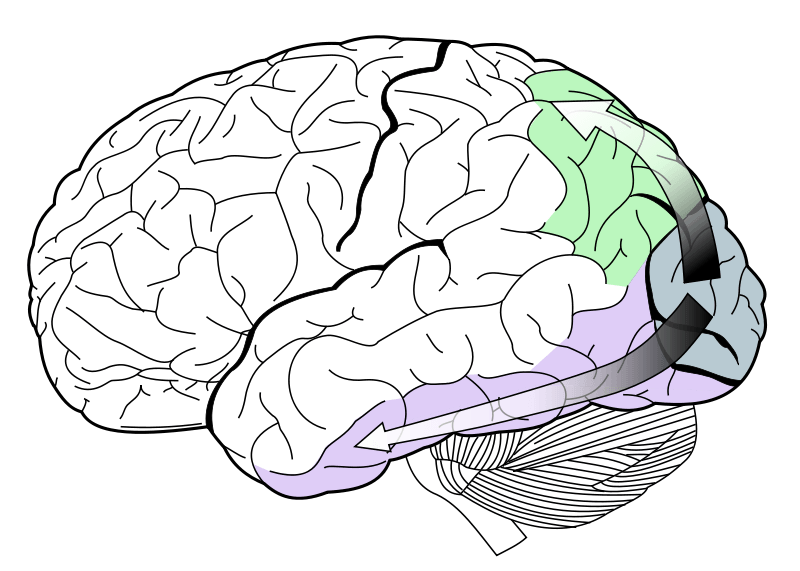
The primary visual cortex, or V1 (shown in teal), is the subject of modeling in the proposed study. The V1 region resides in the occipital lobe and produces the flows of information shown dorsally and ventrally.
Source: Wikipedia
On April 17th, J. Michael Hasse, a PEMM Graduate Student in the Department of Physiology and Neurobiology at the Geisel School of Medicine, gave a presentation at DHMC on his current research project. His research group in Dr. Farran Briggs lab collaborates with Thayer Professor Solomon Diamond and several undergraduates.
The group works together to analyze how external factors affect the physiology of neurons in the primary visual cortex of the brain’s occipital lobe, abbreviated as V1. Ferrets were used for the study because the layout of their flow of visual information to V1 is effectively the same as in humans.
When perceiving visual information, there are a variety of qualities neurons respond to, such as contrast (the difference between black and white) along with spacial frequency and size. To limit the experimental variables, scientists will typically monitor the physiological responses in the brain to fluctuating bars of black and white in experiments with the perception of contrast. They change the width, orientation, and the rate at which the bars cycle. Based on this information many computational models have been made for simple receptive fields that can predict responses to stimuli.
With more complex visual stimuli, these simple receptive models fall apart. Real life has far more complex visual patterns; contrasts of colors show us the outline of a tree, not a simple black or white line. Hasse seeks to determine the nature of cognitive difference between viewing these bars and the real world and what the magnitude of that difference is. At this point, no one knows how far off these models are from representing true natural vision.
The experiment is simple and engaging: they will monitor the differences in how a ferret perceives visual stimuli from a natural environment and compare it to perception of a digital environment. The ferrets are first placed in a pen representing the natural world, covered in images of trees, branches, and leaves. The Diamond lab complied these images using an original algorithm to have similar luminance, so as to limit confound variables. Using motion sensor technology in the form of a surgically implanted array with 36 electrodes and tripods, the ferrets’ experience in the pen is then recreated and transformed into a 3-D movie that the ferrets later watch in an anesthetized state.
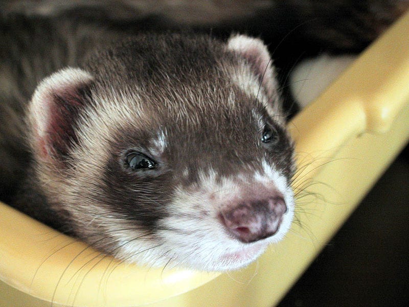
Ferrets were chosen for the study in part due to the similarity of their neural pathways involved in visual perception to those of humans.
Source: Wikimedia
There are two questions this experiment will immediately seek to analyze. First, are receptive fields different for animals exposed to the natural stimuli when compared to those exposed to the simulation? To answer this the experiment will quantify differences in shape and tuning responses of receptive fields between the two conditions – natural and digital. Second, can a computational model be created to better predict responses in natural conditions? The group will compile the data and find the average stimulus to give an idea of the linear receptive field and the tuning properties of the cell.
In the question and answer session after the presentation, audience members made two interesting critiques. First, the ferrets have never experienced life outside of captivity, as they were raised in labs for experimentation. How do we know these natural images mean the same to them as to an animal from the wild? The neurons in their brains could have developed differently in captivity. Secondly, is there a difference in the ferret’s neuron receptivity when it is awake wandering the pen versus under anesthetic watching the 3D video? The anesthetic may have some sort of effect on this perception.
Michael Hasse is coming close to connecting the gap between simple experiments and the real, natural world – one of the largest problems of visual physiology.